Background: Paroxysmal nocturnal hemoglobinuria (PNH) is a rare clonogenic disease of hematopoietic stem cells. LncRNAs has a wide range of biological functions, including cell differentiation, cell proliferation and substance metabolism. LncRNAs maybe contribute to the proliferation of PNH clones.
Methods: CD59- and CD59+ granulocytes and monocytes cells were sorted by FCM and analyzed by RNA sequencing in 5 PNH patients. We focus on the proliferation relative pathway-NF-κB pathway. The mRNAs which FPKM>10 and over 3 patients were chosen to search out the upstream regulation LncRNAs. Then the expression of LncRNAs were detected by qRT-PCR in 30 PNH patients. The highly expressed LncRNA FAM157C was screened out, and analyzed the correlation with clinical index. Finally, we knock-down FAM157C gene in the PIGA knocked out THP-1 cells by lentivirus transfection technique, and observe the cell proliferation, apoptosis to verify its function.
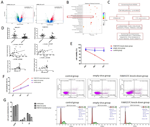
Results: Transcription analysis revealed that 742 upregulation LncRNAs and 3276 upregulation mRNAs were identified in CD59- cells (Figure A). The highly expressed NF-κB pathway (Figure B) mRNAs were analysed by co-expression, after that MALAT1, LINC01002, FAM157C, CTD-2530H12.2, XLOC-064331 and XLOC-106677 were concerned with the 8 mRNAs (Figure C). The results showed that the levels of MALAT1 and FAM157C in CD59- cells expression were significantly higher than that of the CD59+ cell in 30 PNH patients (p<0.05). The expression level of MALAT1 and FAM157C were positive correlation with LDH level and CD59- granulated and monocytes cells ratio (Figure D).
Lentivirus FAM157C transfection knock-down FAM157C gene expression (90%) in the PIGA knocked out THP-1 cells. The cell proliferation assay results showed that there was no significant change in the cell viability at 24h after transfection. But with the transfection time, the cell proliferation activity showed a decreasing trend. The cell viability of the control group, empty virus group and FAM157C knock-down group were (100±0), (93.75±5.995), (77.49±6.597) and (100±0), (92.795±5.802), (60.47±2.059) after 48h, 72h transfection respectively (p=0.0069, 0.0002) (Figure E). The apoptosis rate of control group, empty virus group and FAM157C knock-down group were (2.483±0.3083)%, (2.926±0.5517)%, (6.256±0.5453)% and (5.593±0.6400)%, (6.723±0.3256)%, (11.30±1.075)% and (9.797±0.3235)%, (10.21±0.3005)%, (18.81±0.5363)% after 24h, 48h, 72h transfection respectively (p=0.0006, 0.0005, < 0.0001). The cell apoptosis experiment showed that apoptosis rate increased after transfection of lentivirus FAM157C (Figure F). The results of cell cycle test showed that the G0/G1 phase of the control group, empty virus group and FAM157C knockdown group were (62.98±1.513)%, (65.95±1.174)% and (70.00±0.2404)%, S phase were (3.825±0.7849)%, (5.920±0.9192)% and (13.47±1.039)%, G2 phase were (32.81±1.612)%, (27.47±1.160)% and (16.54±0.7990)% after transfection of lentivirus FAM157C (p=0.0269, 0.0198, 0.0145) (Figure G).
Conclusion: High expressed FAM157C was associated with hemolysis index in PNH, and knock-down it can decrease proliferation ability, induce the apoptosis and the cells were blocked in G0/G1 phase and S phase, indicating FAM157C may be involved in the proliferation of PNH clones.
Key words: Paroxysmal Nocturnal Hemoglobinuria, LncRNAs, clone proliferation NF-κB pathway, LncRNA FAM157C
Figure Legends
Figure A: Volcanic map of differentially expressed LncRNAs and mRNAs, C2 represents CD59- cells, and C1 represents CD59+ cells.
Figure B: Scatter plot is a graphical representation of KEGG enrichment analysis results.
Figure C: Screening of mRNAs and LncRNAs from NF-κB pathway.
Figure D: Correlation analysis between MALAT1 and FAM157C expression and clinical date.
Figure E: The cell proliferation assay was examined by CCK-8 kit.
Figure F: The Cell apoptosis rate was examined by flow cytometry.
Figure G: After FAM157C knockdown, the proportion of cells in G0/G1 phase and S phase increased, while the proportion of cells in G2 phase decreased, and the cells were blocked in G0/G1 phase and S phase.
No relevant conflicts of interest to declare.
Author notes
Asterisk with author names denotes non-ASH members.


This feature is available to Subscribers Only
Sign In or Create an Account Close Modal